-
 The PET scatter phantom is an acceptance testing tool used to determine the imaging systems relative sensitivity to scatter radiation. It can be used to measure the effects of dead-time and the effects of random events generated at different levels of activity of the line source.
 -
 The Nema Scatter Phantom is designed in accordance with the recommendations by the National Electrical Manufacturers Association (NEMA) to standardize the measurement of count rate performance of a scintillation camera in the presence of scatter.
 -
 The NEMA SPECT Triple Line Source Phantom is designed in accordance with the recommendations by the National Electrical manufacturers Association (NEMA) to standardize the measurement of reconstructed spatial resolution of SPECT
 -
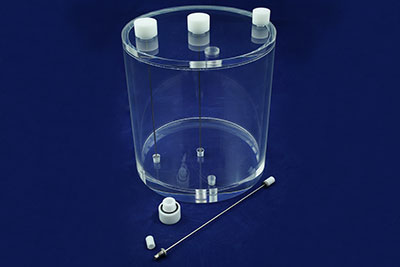
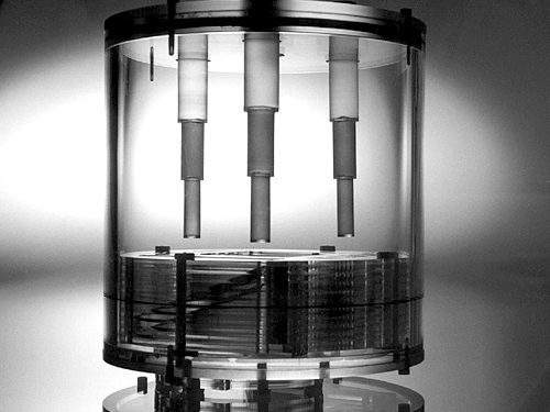
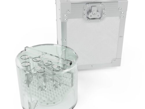
 ECT Line Source Phantom (SMR355) The ECT Line Source Phantom is a cylindrical, liquid filled container with 3 line source inserts.
 -
 This module can be used as a standalone in air or in water if mounted in the Pro-NM Performance cylinder. It can be used to evaluate changes of radius-of rotation on spatial resolution, spatial resolution measurement in air and in water, quantitative evaluation of reconstruction filters and scatter compensation methods.
 -
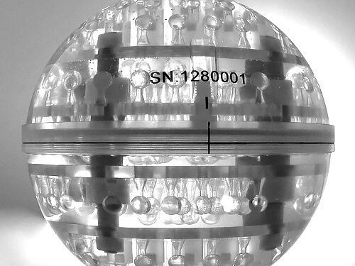
 A phantom for evaluation of spatial resolution of positron emission tomographs (PET). It is used to characterize the widths of the reconstructed image point spread functions (PSF) of compact radioactive sources. It has been designed in accordance with the NEMA Nu 2-2012.
 -
 This SPECT dedicated phantom can be used to produce images similar to those obtained in a whole body tomographic imaging study, with hot and cold lesions and a large cold region representing the lung.
 -
 The phantom for NM systems performance evaluation: routine quality assurance tests, as well as extensive acceptance tests. It can be used to evaluate: pixel size, spatial linearity, RMS noise, signal to noise ratio (SNR), slice width, uniformity, spatial resolution, point spread function, slice position verification, slice incrementation, accuracy, center of rotation, verification, volume sensitivity and low contrast sensitivity.
 -
 The ECTphan™ Phantom is a cylindrical, liquid filled container with a variety of tests for quality monitoring of single photon emission tomography (SPECT) systems .
 -
 The ECTphan™ Phantom is a cylindrical, liquid filled container with a variety of tests for quality monitoring of single photon emission tomography (SPECT) systems.
 -
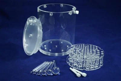 The Small Flangeless Jaszczak SPECT Phantom is used with high spatial resolution SPECT systems. The phantom has a cylinder Twist and Lock lid design.
 -
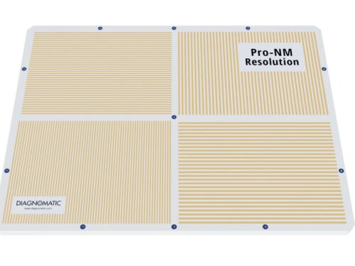 The bar phantom for determination of resolution of Scintillation Cameras. Four-quadrant phantom offers precise determination of camera intrinsic resolution, collimator spatial resolution, field size and linearity.
 -
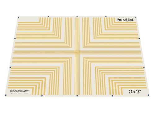 Pro-NM ResL for determination of resolution of Scintillation Cameras. The phantom offers precise determination of camera intrinsic resolution, collimator spatial resolution, field size and linearity.
 -
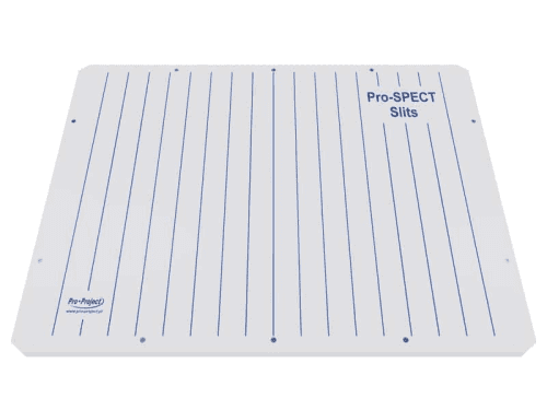 Slits phantom for Intrinsic Spatial Resolution evaluation (Quantitative technique) according to NEMA Standards Publication NU 1-2012. In addition to this standard size phantom, we offer different sizes and configurations manufactured to the highest quality standards.
 -
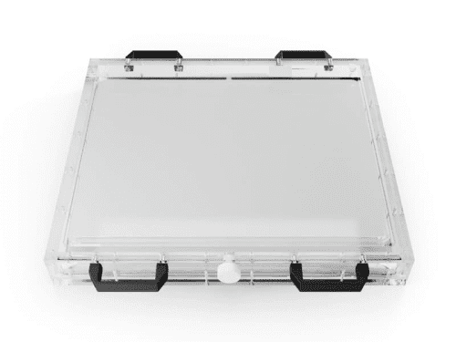 The Pro-NM AutoFlood is dedicated for weekly inhomogeneity and sensitivity control and acquisition of correction matrices, according to revised Guideline on Radiation Protection in Medicine.
 -
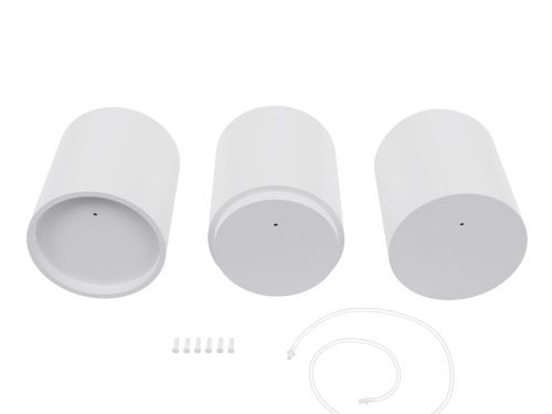
 Flood phantoms provide a simple and efficient means of obtaining optimum camera performance with respect to uniformity of response over the entire crystal area. These phantoms are designed to be filled in horizontal position, thus preventing slight bulging caused by water pressure during vertical filling. Therefore, better uniformity in distribution of activity can be achieved.
 -
 Flood phantoms provide a simple and efficient means of obtaining optimum camera performance with respect to uniformity of response over the entire crystal area.
 -
 A simple phantom the quality assurance of geometric distortion and spatial resolution of gamma cameras. Array of holes, which when filled with activity, allows to measure point-to-point distances and Point Spread Function (PSF) - spatial resolution - at each point and its homogeneity across the entire Field of View.
 -
 This scatter phantom simulates in-vivo forward and backscatter characteristics of 99mTc gamma rays for the extrinsic measurement of a scintillation camera’s deadtime. The phantom produces a spectrum typical of that observed from 99mTc in the myocardium. Reference: Ralph Adams, Gerald J. Hine, and C. Duane Zimmerman, “Deadtime Measurements in Scintillation Cameras Under Scatter Conditions Simulating Quantitative Nuclear Cardiography,” The Journal of Nuclear Medicine, 19 (1978), 538-544.
 -
 LiquiPhil™ Phantoms are excellent for calibration, quality assurance, research, and training in nuclear medicine and SPECT studies. The phantoms are constructed of cellulose acetate butyrate, a transparent material that provides strength and low water absorption.
 -
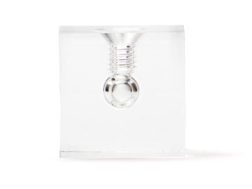 A set of 7 clear PMMA cubes, 17x17x17mm, with a 5mm dia. spherical fillable void, which can be filled with any mixture of radionuclide, CT contrast agent or MR contrast agent and sealed with a nylon screw (provided).
 -
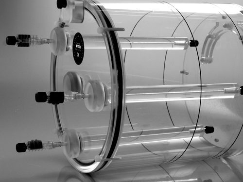
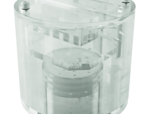
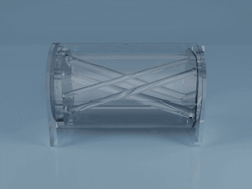 The Phantom designed for a simple and cost effective verification of image alignment and distortion in hybrid scanning systems like PET/CT or NM/CT. It consists of a cylinder that can be filled with a variety of fluids. Several non-parallel rods of varying diameter and at certain angles in relation to the phantom’s axes run the entire length of the cylinder.
 -
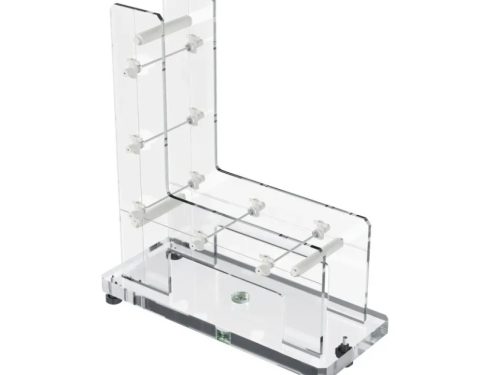
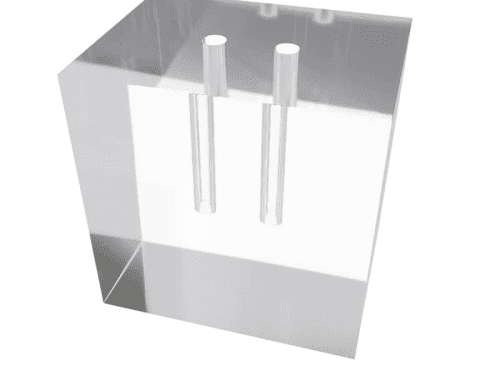
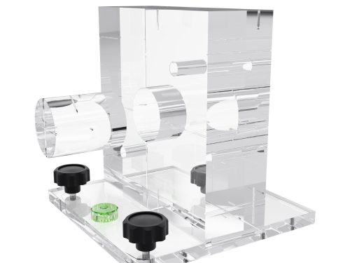
 The 3D Image Fusion Ramp Cube enables verification of registration for SPECT/CT and PET/CT fusion imaging.
 -
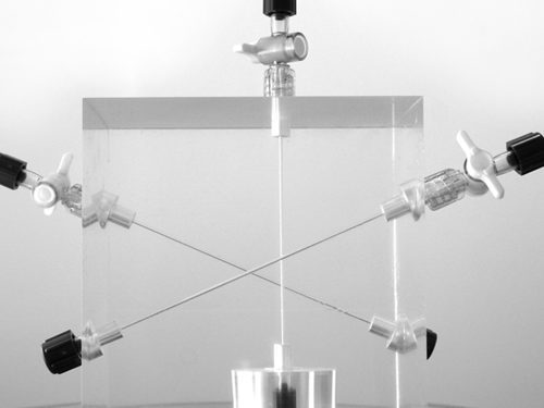
 This phantom can be used to check the alignment of the internal and external lasers to the radiographic centre of CT and PET/CT units and to verify lateral gantry angle. It can also be used with accelerator units to check vertical and lateral gantry angles, laser alignment and vertical table movement.
 -
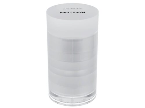 A compact CT phantom designed for small-animal imaging with full clinical-grade QA functionality.
 -
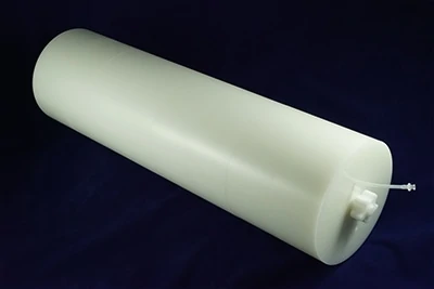
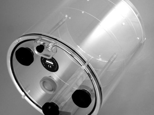
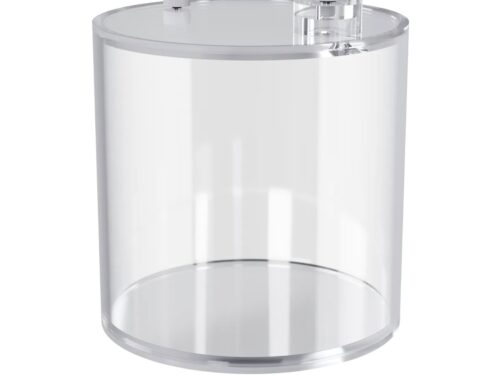 Cylindrical water phantom with a dedicated infusion spot. The cylinder can be used to examine reconstructed images (tomography slices) for uniformity, artifacts, activity concentration, and similar purposes.
 -
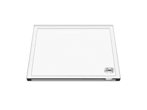
 ACR accredited Medium MRI phantom for comprehensive evaluation of critical imaging parameters of magnetic resonance imaging (MRI) in a time efficient manner. The phantom can be used for the measurement of absolute values for calibration purposes. However, its design is optimized for time efficient daily quality assurance too.
 -
Large MRI phantom for comprehensive evaluation of critical imaging parameters of magnetic resonance imaging (MRI) in a time efficient manner. The phantom can be used for the measurement of absolute values for calibration purposes. However, its design is optimized for time efficient daily quality assurance too.
-
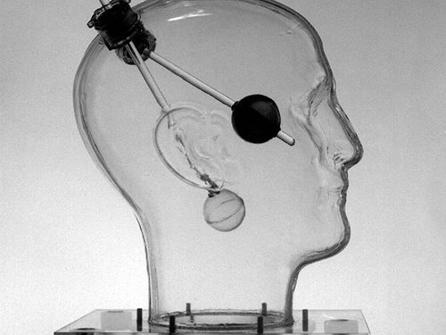
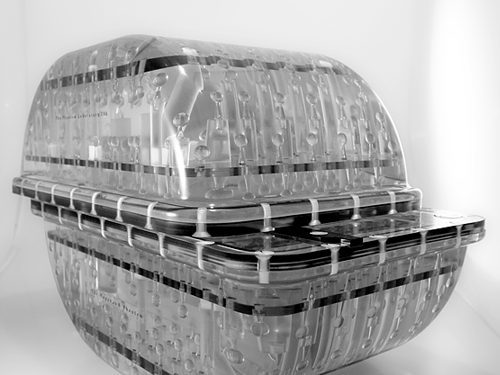 An integrated MR QA system for MR guided surgery and radiotherapy MRI manufacturers have made great strides in reducing MR system distortion. Maintaining acceptable levels of distortion relies on properly controlling a long chain of conditions. It is critical to have a robust system of quality control for key imaging performance characteristics in order to detect significant deviations before they affect clinical operations.
 -
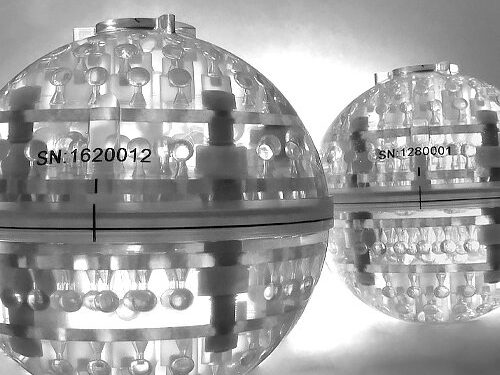 The Magphan® S162 system phantom is designed to meet the specific QA needs for MRI scanners used in quantitative imaging, particularly in applications where geometric distortion can have critical impacts.
 -
 The EMR128 is from the family of Magphan® Quantitative Imaging Phantoms developed with physicist Richard Mallozzi, Ph.D. The Magphan® 128 is designed to provide detailed distortion mapping of MR head coil sized fields.
 -
 Phantom for comprehensive evaluation of critical imaging parameters of magnetic resonance imaging (MRI) in a time efficient manner.
 -
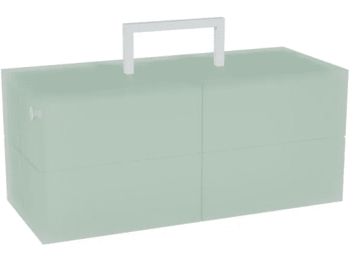 Rectangular MRI phantom simulating attenuation of the human spine.
 -
 This stability (agar) phantom consists of a cylindrical phantom and agar material inside. Using this phantom a Signal to Noise Ratio, Signal Fluctuation to Noise Ratio, drift, and other imaging measures over a 100-volume or 200-volume fMRI scan can be performed. The agar phantom has characteristics similar to the T2 measures of a human head, but provides no change in signal. The T1 and T2 characteristics of the agar phantom at 3T are ~900 ms T1 and 30 ms T2.
 -
 The Pro-Dent DIN 2D is a universal phantom for carrying out constancy and acceptance tests of conventional and digital dental X-ray units (intra-oral, panoramic, and cephalometric).
 -
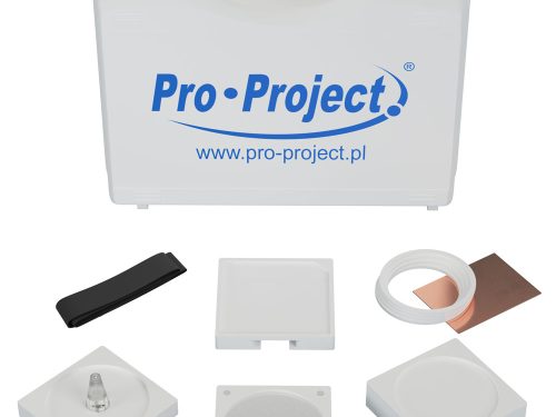 The Pro-Dent set is a universal set of phantoms for carrying out constancy and acceptance tests of conventional and digital dental X-ray units (intra-oral, panoramic and cephalometric).