-
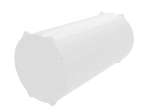
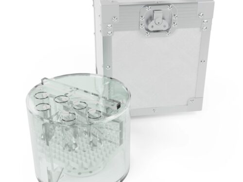
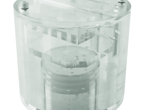
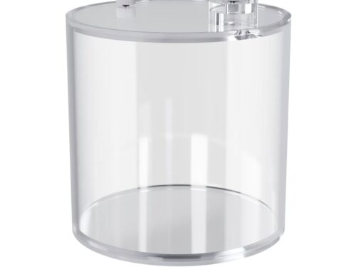 Cylindrical water phantom with a dedicated infusion spot. The cylinder can be used to examine reconstructed images (tomography slices) for uniformity, artifacts, activity concentration, and similar purposes.
 -
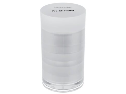 A compact CT phantom designed for small-animal imaging with full clinical-grade QA functionality.
 -
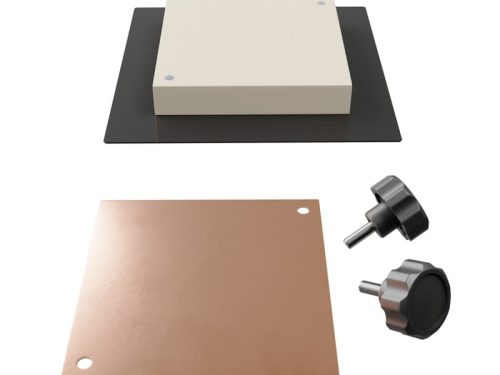
 A set of acrylic plates for testing Automated Exposure Control of radiography equipment.
 -
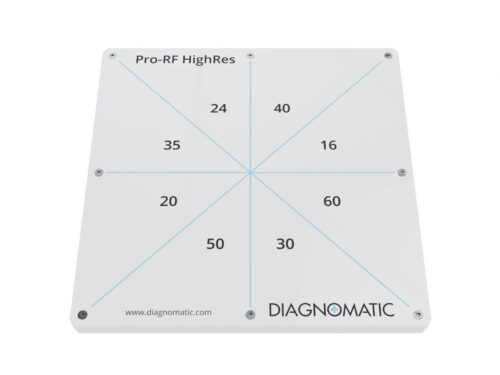 The test phantom for evaluation of high contrast resolution of fluoroscopy system in one exposition. It consists of eight patterns of wire mesh in a pie shape.
 -
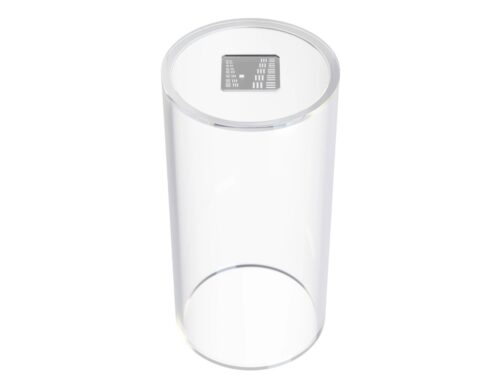 A simple test device for the focal spot size evaluation. It consists of a precision bar pattern placed at the end of the cylinder that provides accurate and reproducible target-to-image receptor spacing.
 -
 Phantom is designed to evaluate digital functions of DSA systems and can be used to check: contrast range, resolution, linearity, uniformity, amplifier dynamic range, registration accuracy and subtraction effectiveness.
 -
 A set of acrylic and aluminium filters for testing diagnostic X-rays according to AAPM recommendations from Report No. 31 ”Standardized Methods for Measuring Diagnostic X-Ray Exposure”.
 -
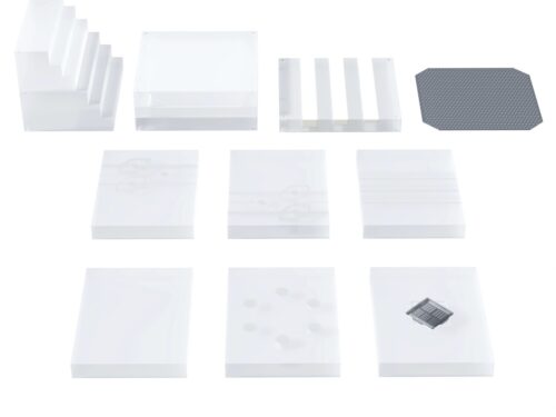
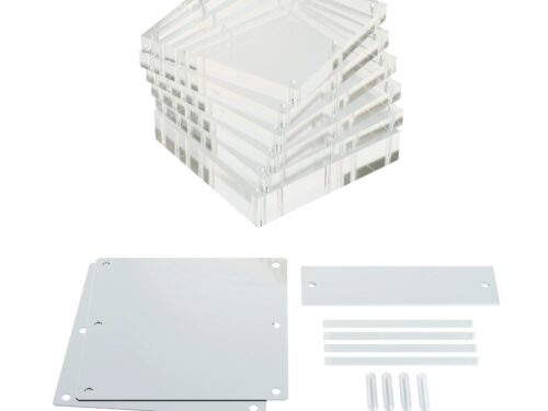
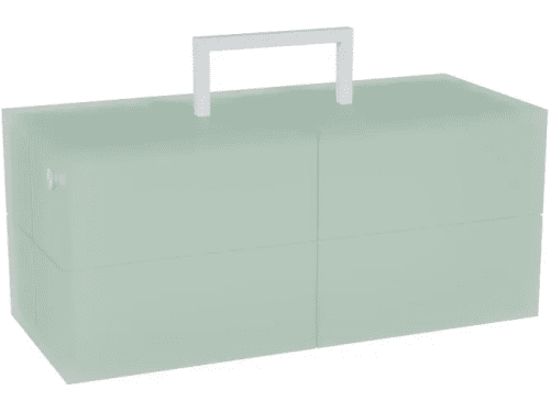
 This modular phantom made of PMMA consists of several plates allowing variation of phantom thickness setup in steps of 25 mm, up to a total of 300 mm, simulating a range of patient sizes.
 -
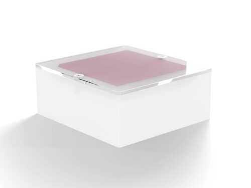
 Ensure superior diagnostic quality of your CBCT system with the Pro-MAM CBCT Phantom. This advanced QA tool enables routine assessment of critical parameters: microcalcification visibility, contrast, resolution, and image uniformity.
 -
 The phantom can test the performance of a mammographic system and its ability to image small structures similar to those found clinically: calcifications, fibrous calcifications in ducts and tumor masses.
 -
 The Pro-Dent DIN 2D is a universal phantom for carrying out constancy and acceptance tests of conventional and digital dental X-ray units (intra-oral, panoramic, and cephalometric).
 -
 This phantom is designed according to ICRU 87 report and AAPM TG-200 specifications.
 -
 A 25 mm aluminum filter with insertion frame for mounting near the X-Ray tube.
 -
 This SPECT dedicated phantom can be used to produce images similar to those obtained in a whole body tomographic imaging study, with hot and cold lesions and a large cold region representing the lung.
 -
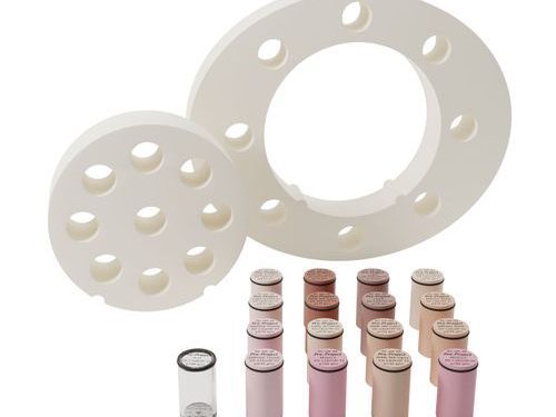 The Pro-RT ED phantom is used to account for tissue heterogeneity in radiotherapy treatment planning. It can be used to define the precise correlation of CT data to the electron density of various tissues. The phantom consists of two nested disks that represent the head and abdomen. Nine different tissue equivalent electron density plugs can be positioned at 17 different locations within the scan field. Included is a water vial plug that can be filled with any fluid. Optional distance marker plugs enable quick assessment of the CT scanner’s distance measurement accuracy.
 -
 Pro-Water Plates makes radiation beam calibration easier than ever. With improved uniformity and durability, Pro-Water Plates enables precise, accurate and effective Quality Assurance. Pro-Water Plates mimics true water within 1% accuracy, supporting accurate calibration for zadiotherapy beams.
 -
 Pro-RT IsoBeam This is a multifunctional, precision quality assurance tool for daily, weekly or monthly quality assessments of all mechanical and geometrical treatment parameters of linear accelerators or teletherapy units. It is designed to easily and accurately check collimator isocentricity, gantry isocentricity, table isocentricity, collimator field size accuracy, radiation/light field congruence, isocenter rotational stability, ODI accuracy and laser light alignments.
 -
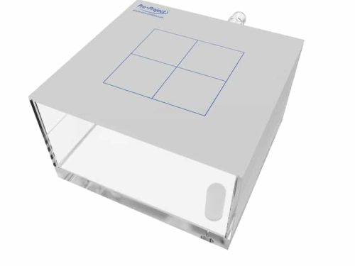
 This phantom is designed for quality control of a CT simulator, which usually consists of a CT scanner, virtual symulator software and a laser marking device (marking the centre of target volume). Since geometrical planning is the core of CT simulation, periodic quality control is essential for maintaining optimum image quality and patient care. The phantom allows performing detailed geometry and table movement tests as well as image quality assessment - low contrast resolution and high contrast detectability.
 -
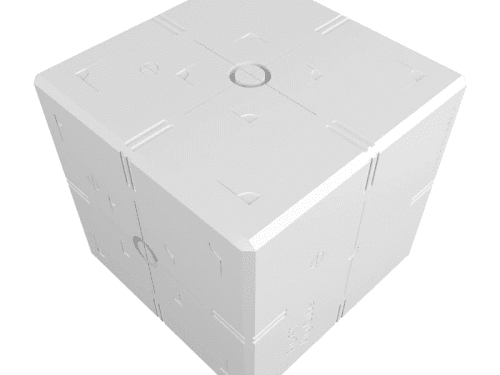 Simple to use and economic tool to perform true spatial alignment and coincidence tests on IGRT systems. Pro-RT Align-Cube contains engraved markings on the exterior and radiographic markers inside. The isocenters of both the OBI and the EPID can be checked for true spatial alignment and coincidence with that of the treatment beam. The phantom ensures the accuracy of linacs’ image-guidance systems, including CBCT, x-ray volumetric imaging (XVI) and on-board imaging (OBI). It can be used to test: 3D cone beam registration kV and MV system coincidence kV and MV projection images laser and light field coincidence weekly remote table adjustments
 -
 The stationary water phantom for high energy photon dosimetry with all types of ionization chambers.
 -
 Phantom for comprehensive evaluation of critical imaging parameters of magnetic resonance imaging (MRI) in a time efficient manner.
 -
 Rectangular MRI phantom simulating attenuation of the human spine.
 -
 This stability (agar) phantom consists of a cylindrical phantom and agar material inside. Using this phantom a Signal to Noise Ratio, Signal Fluctuation to Noise Ratio, drift, and other imaging measures over a 100-volume or 200-volume fMRI scan can be performed. The agar phantom has characteristics similar to the T2 measures of a human head, but provides no change in signal. The T1 and T2 characteristics of the agar phantom at 3T are ~900 ms T1 and 30 ms T2.
 -
 ACR accredited Medium MRI phantom for comprehensive evaluation of critical imaging parameters of magnetic resonance imaging (MRI) in a time efficient manner. The phantom can be used for the measurement of absolute values for calibration purposes. However, its design is optimized for time efficient daily quality assurance too.