-
 White Matter is designed based on average brain anatomy and it is made out of realistic tissue mimicking materials suitable for both Ultrasound and MRI applications. The feature can be incorporated into the brain phantom or simply submerged in a brain mimicking material used to fill the skull or head.
 -
 Tumor for R&D is a perfect example of a feature that can be implemented to any location of the brain or simply submerged in a brain mimicking material used to fill the skull or head phantom.
 -
 Stenosis for R&D is an example of a feature that can be implemented to any location in the brain and/or blood vessels.
 -
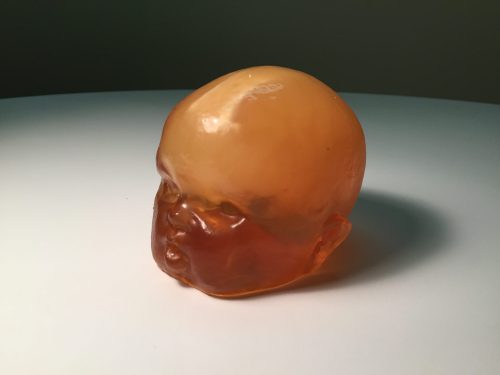
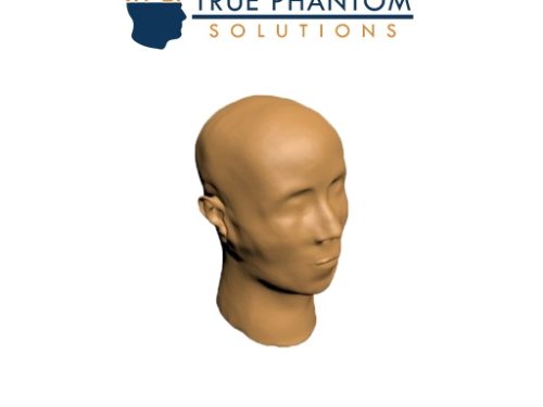 Head skin is designed based on average anatomy of and adult human head. It is made from realistic tissue mimicking material suitable for Ultrasound, MRI and CT applications.
 -
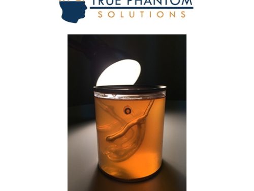
 Blood Vessels for R&D can be fabricated as a stand-alone phantom or a feature implemented to any location of the brain. They can be also simply submerged in a brain mimicking material used to fill the skull and/or head phantom.
 -
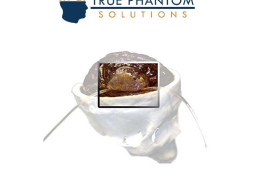 Our Adult Head (Hematoma) phantom is a cutting-edge medical training solution that seamlessly integrates with X-Ray CT, US, and MRI. This advanced phantom replicates the intricate anatomy of the human head, providing healthcare professionals with a realistic and immersive tool for skill development. This phantom is an excellent tool for applications relating to head trauma, hematomas, stroke, aneurysm, etc.
 -
 Foreign Objects for R&D Various foreign objects can be incorporated within the skull directly into the brain tissue or to any other location of the phantom. The objects can mimic different materials such as bullets, shrapnel, and pieces of bones.
 -
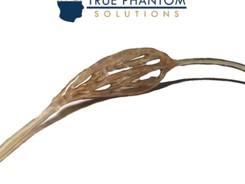
 Falx Cerebri is a membrane which divides left and right brain hemispheres and it is designed based on an average anatomy of an adult human brain. This feature is a perfect navigation point for medical brain imaging and it is made from a realistic material suitable for ultrasound and MRI applications.
 -
 Complex Blood Vessels for R&D is a feature that can be implemented to any location of the brain or simply submerged in a brain mimicking material used to fill the skull or head phantom.
 -
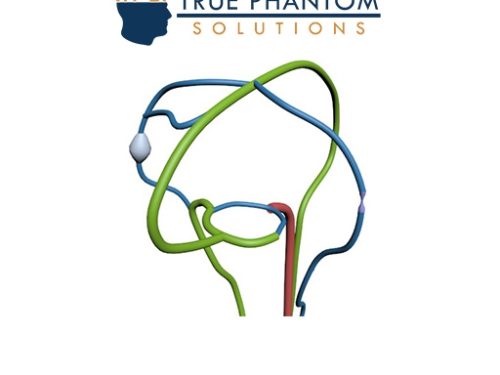 Brain blood vessel feature is designed based on the arterial brain vessels and it is made out of realistic tissue mimicking material suitable for Ultrasound, MRI and CT applications.
 -
 Brain Ventricles for R&D are designed based on an average anatomy of human brain ventricles and they are fabricated using a novel 3D printing technology.
 -
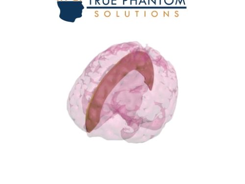
 The brain structure is created based on an average anatomy and dimensions of an adult brain. The parenchyma is be made out of a homogeneous material with customizable composition to suit the need of various biomedical projects.
 -
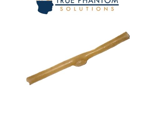
 Bifurcation for R&D is a feature which can be implemented to any location of the brain/blood vessels or simply submerged in a brain mimicking material used to fill the skull or head phantom.
 -
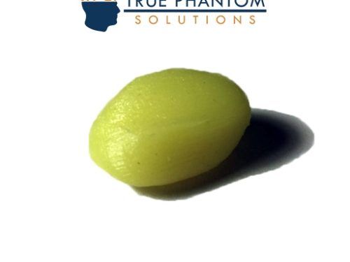
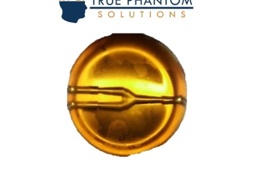
 Aneurysm for R&D is an example of a feature that can be implemented to any location in the brain and/or blood vessels.
 -
 Sectional Mouse Phantom for X-Ray/CT is a phantom designed based off of the average anatomy of a mouse. It is sliced into segments and is used for medical imaging research and development, testing, and calibration-related work.
 -
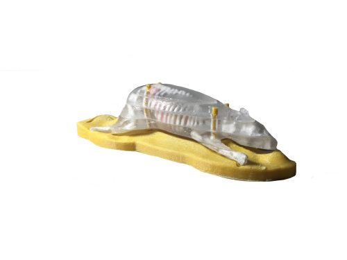
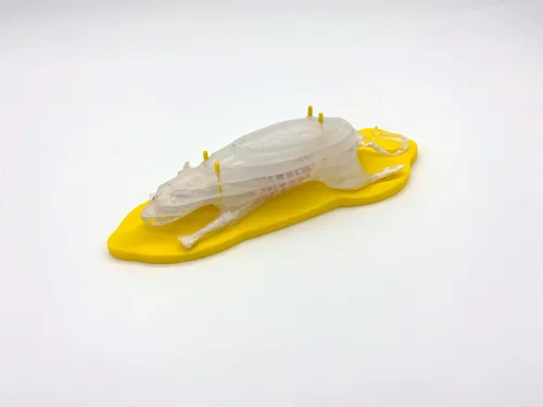 Sectional Rat Phantom for X-Ray/CT is a phantom designed based off of the average anatomy of a rat. It is sliced into segments and is used for medical imaging research and development, testing, and calibration-related work.
 -
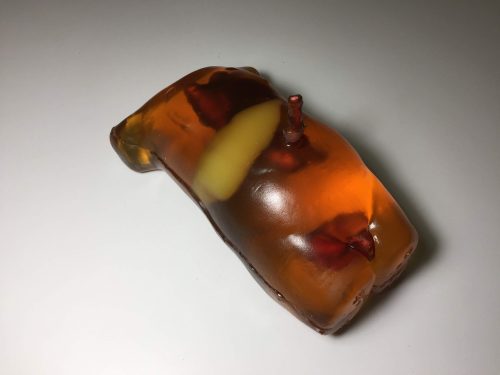
 Rat Phantom (Cylindrical) can be fabricated as a simple cylinder or as an anatomically correct rat. Either of these phantoms is a powerful tool that can be used for testing and calibration-related work of various medical imaging devices.
 -
 Rat Phantom (Cylindrical) can be fabricated as a simple cylinder or as an anatomically correct rat. Either of these phantoms is a powerful tool that can be used for testing and calibration-related work of various medical imaging devices.
 -
 The Rat Phantom is built to resemble the average anatomy of a rat or mouse. It is compatible with X-Ray/CT and MRI imaging methods. Customizable with extra chambers and features, it is a versatile tool for bio-medical research projects and can be used for testing and calibration-related work.
 -
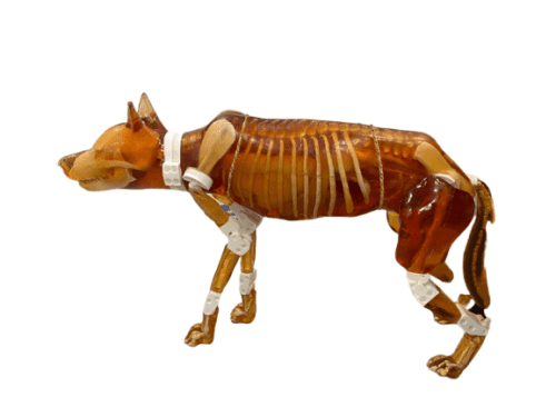 The newly designed detachable dog phantom serves as an independent training simulator compatible with ultrasound and X-Ray/CT imaging. It is an ideal teaching tool for veterinary professionals, featuring improved anatomical structures and removable body parts. This allows for practicing various positioning techniques under both imaging modalities.
 -
 The Newborn Torso (Transparent) phantom was developed in collaboration with the Sonographic Clinical Assessment of the Newborn Training Program at the University of Calgary. This phantom is an deal tool for Ultrasound-guided procedures, such as catheter insertion through the umbilical cord and bladder catheterization. It is constructed with realistic tissue-mimicking materials suitable for Ultrasound and X-Ray/CT imaging.
 -
 The Newborn Torso phantom was developed in collaboration with the Sonographic Clinical Assessment of the Newborn Training Program at the University of Calgary. This phantom is an deal tool for Ultrasound-guided procedures, such as catheter insertion through the umbilical cord and bladder catheterization. It is constructed with realistic tissue-mimicking materials suitable for Ultrasound and X-Ray/CT imaging.
 -
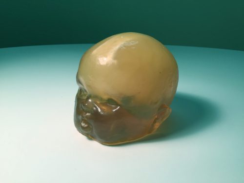 The Newborn Head (Simple) phantom is designed based on an average newborn head and is made out of realistic tissue-mimicking materials suitable for Ultrasound imaging.
 -
 The Newborn Head (Complex) Phantom is a training tool for diagnostic medical imaging and POCUS. Based on an average newborn head, it is made from realistic tissue-mimicking materials suitable for Ultrasound and X-Ray/CT imaging.