-
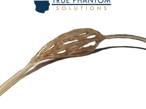
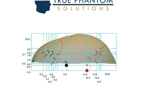
 The Newborn Torso (Transparent) phantom was developed in collaboration with the Sonographic Clinical Assessment of the Newborn Training Program at the University of Calgary. This phantom is an deal tool for Ultrasound-guided procedures, such as catheter insertion through the umbilical cord and bladder catheterization. It is constructed with realistic tissue-mimicking materials suitable for Ultrasound and X-Ray/CT imaging.
 -
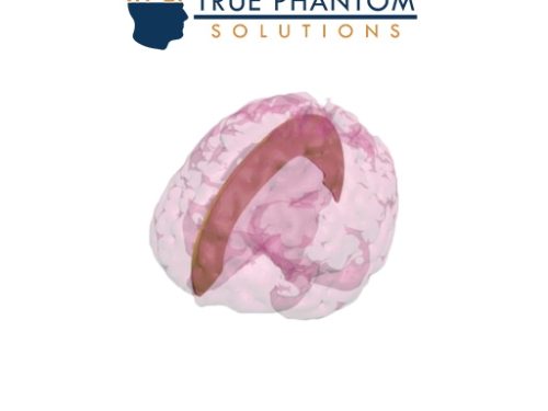 Falx Cerebri is a membrane which divides left and right brain hemispheres and it is designed based on an average anatomy of an adult human brain. This feature is a perfect navigation point for medical brain imaging and it is made from a realistic material suitable for ultrasound and MRI applications.
 -
 The Pediatric Full Body Phantom is an X-Ray/CT and Ultrasound-compatible training product used for training patient positioning techniques. It is popular among medical schools and teaching hospitals for training radiology students and medical professionals. This life-size phantom consists of anatomically correct organs and bones divided into 10 body parts.
 -
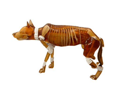 The newly designed detachable dog phantom serves as an independent training simulator compatible with ultrasound and X-Ray/CT imaging. It is an ideal teaching tool for veterinary professionals, featuring improved anatomical structures and removable body parts. This allows for practicing various positioning techniques under both imaging modalities.
 -
 The dog phantom is a versatile training simulator for sonographers, radiographers, and veterinary professionals. Compatible with X-Ray/CT and MRI imaging, it offers improved anatomical structures and removable body parts, allowing for practice in different positioning techniques. An ideal teaching tool with independence from external hardware/software.
 -
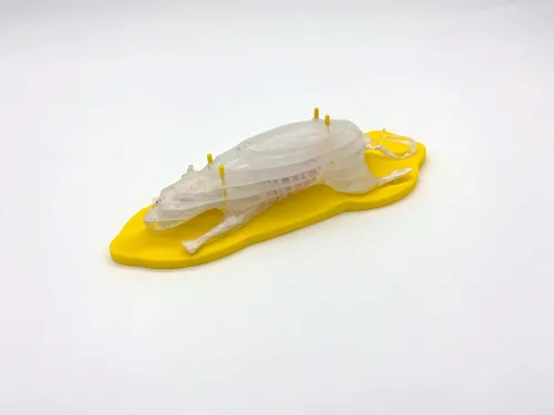
 Rat Phantom (Anatomical) is based on the average anatomy of a rat/mouse and is suitable for both X-Ray/CT and MR imaging methods, and it can be customized to excel in a selected imaging modality. It is a powerful tool that can be used for testing and calibration-related work of various medical imaging devices.

