-
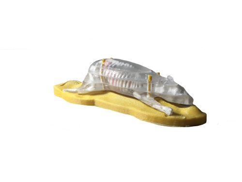 Sectional Mouse Phantom for X-Ray/CT is a phantom designed based off of the average anatomy of a mouse. It is sliced into segments and is used for medical imaging research and development, testing, and calibration-related work.
 -
 The Advanced Newborn Torso phantom closely resembles the anatomy of an average-sized newborn torso. It is constructed with realistic tissue-mimicking materials suitable for MRI and X-Ray/CT imaging. The phantom features a water-fillable umbilical cord and bladder, facilitating catheterization training.
 -
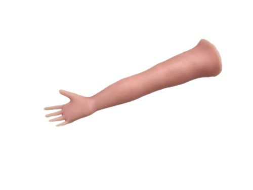 The Newborn Arm Phantom is designed to resemble the anatomy of a healthy newborn baby’s arm. Made from realistic materials, it is suitable for ultrasound-guided IV training for healthcare professionals. This durable and long-lasting phantom allows for practice in intravenous injections under ultrasound guidance, with permanently prefilled blood vessels, eliminating the need for additional fluid.
 -
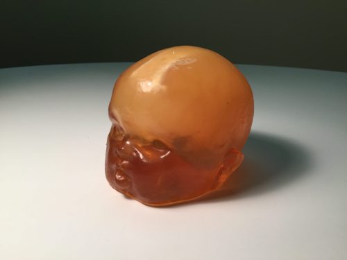
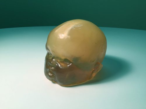
 The Adult Human Skull with Brain Phantom is a versatile tool ideal for medical research and treatment planning, specifically for non-invasive HIFU brain surgeries and medical imaging studies. Resembling the Head Phantom but without skin-mimicking tissue, this model features a realistic three-layered structure with an inner diploe layer, closely resembling the average human male skull. Crafted from a patented epoxy-based bone material, it ensures compatibility with Ultrasound, MRI, and X-Ray/CT applications. Customization options for different parts of the skull are also available upon request. The phantom incorporates the complete ventricular system, including lateral, third, and fourth ventricles. These inflatable ventricles simulate realistic pressure dynamics within the brain, closely approximating actual cerebral anatomy. The ventricles can be filled with various liquids to achieve desired effects. For MRI applications, the phantom’s tissues exhibit realistic T2 relaxation time values, making it an excellent fit for T2-weighted MRI imaging techniques. Proton Density imaging methods also yield high-quality outcomes. While T1-weighted techniques are viable, T1 values are less accurate, falling within the approximate range of 100 ms. Before imaging the phantom, please fill the ventricles with water (or chosen contrast agent) using the tube on the back of the brain. After filling the ventricles, plug the tube with a removable blue silicone plug.
 -
 The Newborn Torso phantom was developed in collaboration with the Sonographic Clinical Assessment of the Newborn Training Program at the University of Calgary. This phantom is an deal tool for Ultrasound-guided procedures, such as catheter insertion through the umbilical cord and bladder catheterization. It is constructed with realistic tissue-mimicking materials suitable for Ultrasound and X-Ray/CT imaging.
 -
 Our Adult Spine phantom consists of 25 individual vertebrae assembled on top of each other and encased in a semi-cylindrical-shaped body-mimicking tissue. It is majorly composed of the following components.
 -
 The Newborn Head (Complex) Phantom is a training tool for diagnostic medical imaging and POCUS. Based on an average newborn head, it is made from realistic tissue-mimicking materials suitable for Ultrasound and X-Ray/CT imaging.
 -
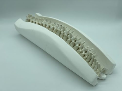 The Adult Spine Phantom comprises 25 vertebrae arranged in a semi-cylindrical body-mimicking tissue. The vertebrae have a realistic three-layered structure with adjustable inner porosity. Made from patented bone material suitable for X-Ray/CT, it is used in medical imaging research and treatment planning.
 -
 The Newborn Head (Simple) phantom is designed based on an average newborn head and is made out of realistic tissue-mimicking materials suitable for Ultrasound imaging.
 -
The Adult Spine Phantom comprises 25 vertebrae arranged in a semi-cylindrical body-mimicking tissue. The vertebrae have a realistic three-layered structure with adjustable inner porosity. Made from patented bone material suitable for Ultrasound and X-Ray/CT, it is used in medical imaging research and treatment planning.
-
 Doppler Phantom serves purposes in medical imaging research, development, training, and testing. The phantom includes a bone plate simulating the 2D projection of the skull with realistic internal porosity. Internal blood vessels can be filled with water or commonly used blood-mimicking fluids.
 -
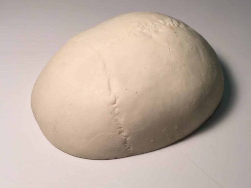 The Standard Calvaria phantom is an excellent tool for medical imaging research. The skull bones are made from a realistic patented epoxy-based bone material suitable for Ultrasound, MRI, and X-Ray/CT applications.