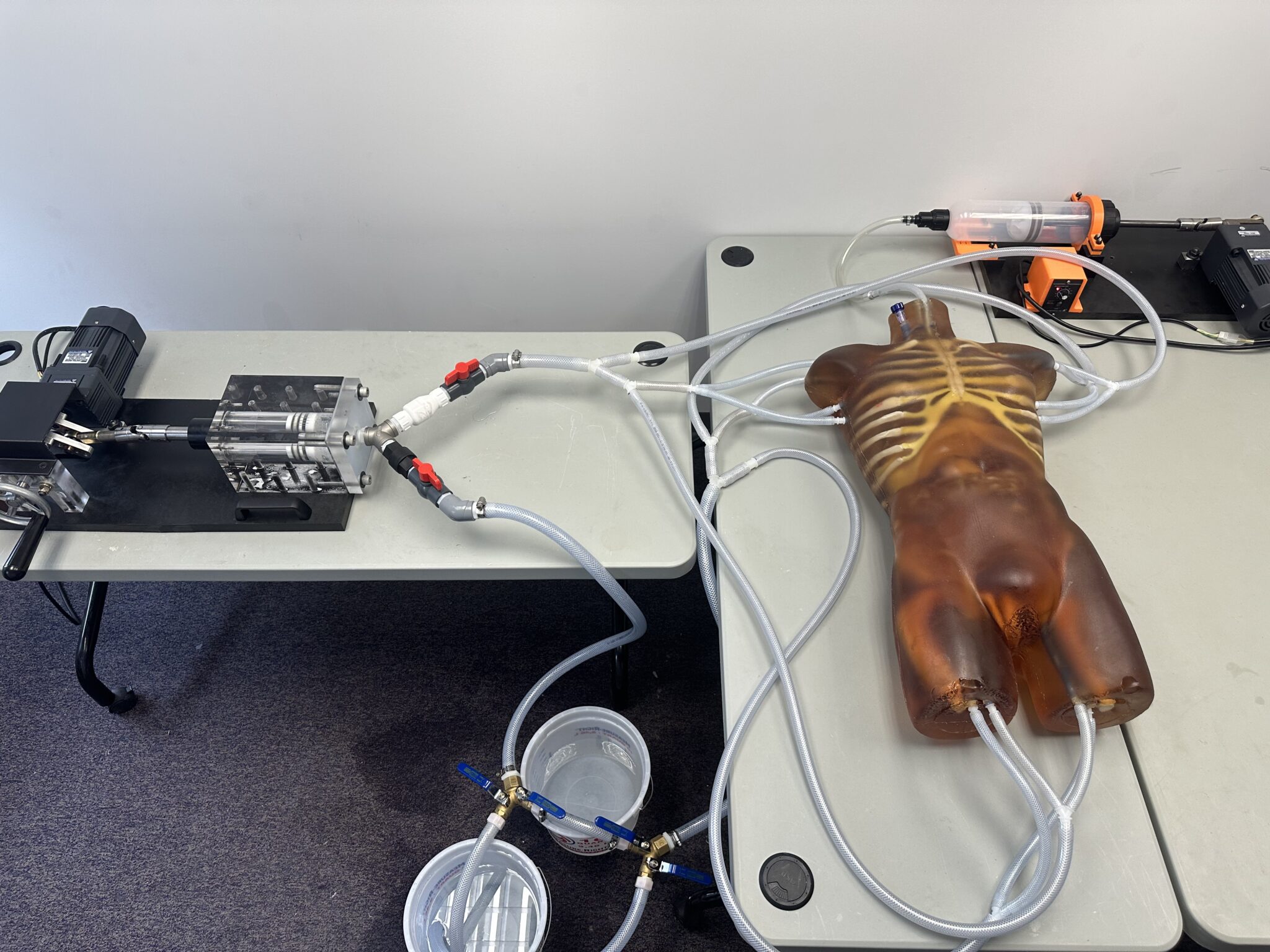
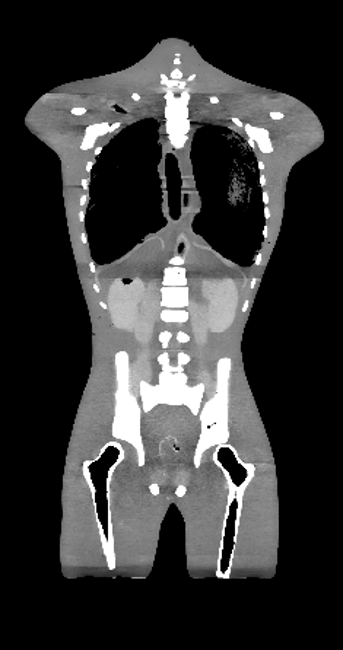
Adult Human Torso (Dynamic) with a beating heart and breathable lungs is a lifelike, anatomically realistic, from neck to pelvic area phantom/simulator with all the essential organs present inside. This phantom is compatible with MRI and X-Ray/CT, and it is an ideal tool for applications such as angiography. The organs are made from realistic urethane-based tissue-mimicking materials, and the bones are made from a patented ceramic-reinforced epoxy-based composite material. The skeleton is assembled from individually cast bones with vertebrae that have a realistic three-layered structure and inner porosity.
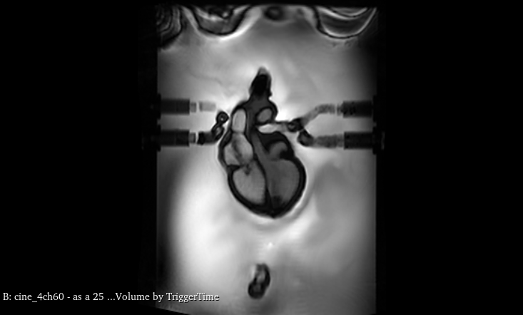
In terms of MRI applications, the phantom tissues have realistic T2 relaxation time values, which makes this product to be the best fit for any T2-weighted MRI imaging methods. Very good results can also be achieved with Proton Density imaging methods. The phantom can still be imaged with T1-weighted methods, but the T1 values are less realistic, and they are within the range of about 100 ms.
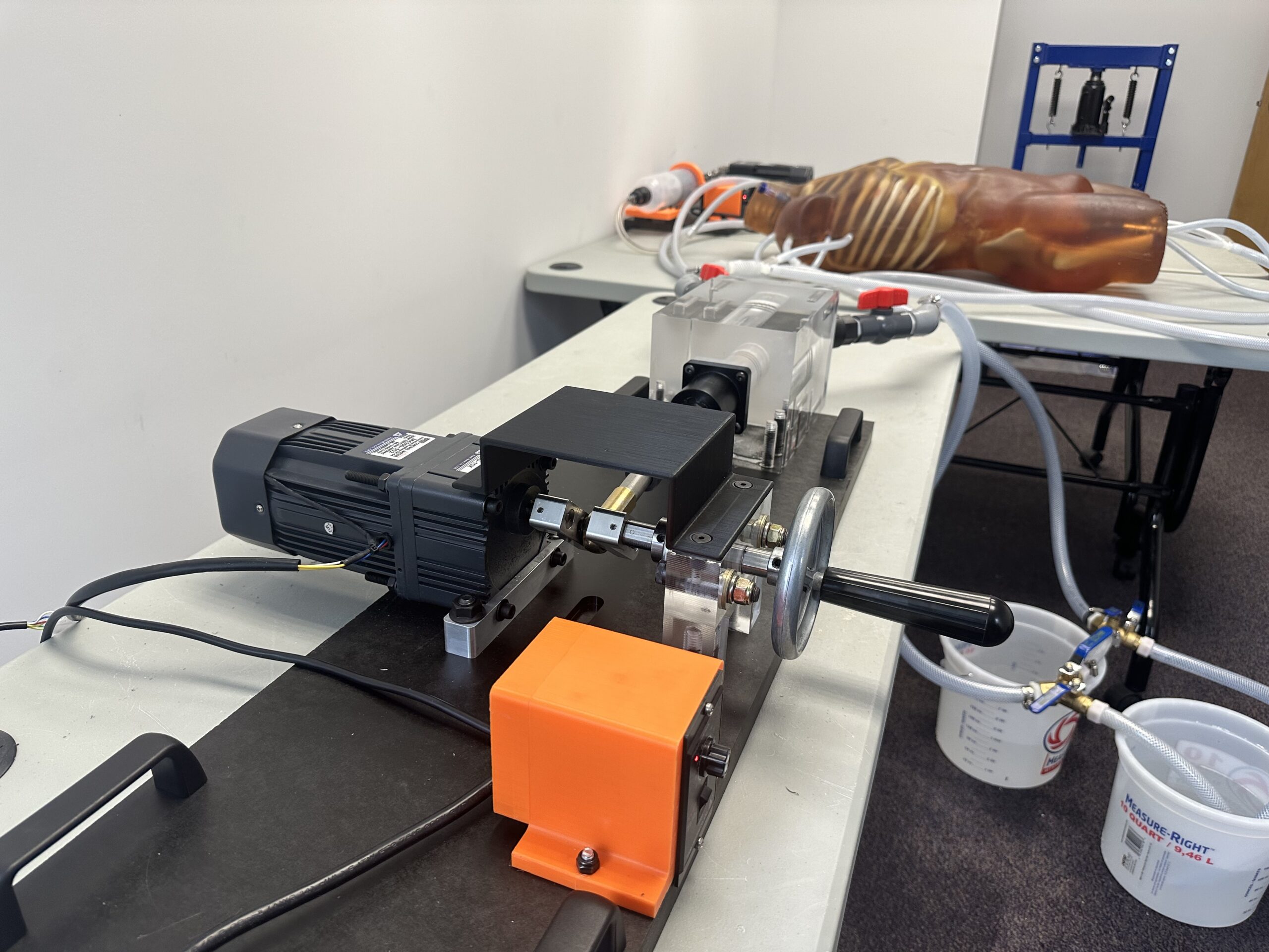
The design of this phantom includes a realistic heart with all major heart vessels and internal heart chambers. The TPS heart pump is included with this phantom, and it enables the realistic heartbeat and motion of heart walls. The TPS lung pump is also included to provide lifelike breathing motion in the torso. https://youtu.be/E24hHyoGwYI
Anatomy:
Complete Spine Complete Ribcage Shoulders and Clavicles Pelvis Partial Femoral Bones Trachea Realistic Beating Heart with:
Complete Spine
Complete Ribcage
Shoulders and Clavicles
Pelvis
Partial Femoral Bones
Trachea
Realistic Beating Heart with:
– 2 Atria, 2 Ventricles:
– Right Ventricle
– Moderator Band that runs from the anterior wall to the septum bridge
– Left Ventricle
– Papillary muscles to the anterior and posterior walls
– Right Atrium
– Right atrial appendage with distinct muscle structures
– Left Atrium
– Unique muscle structure to the appendage of the left atrium
Appendage Feature
Aorta
Jugular vessels
Pulmonic and Pulmonary Veins attached to LA
Flexible Valvular Structures
SVC
IVC
Inflatable Breathing Lungs
Diaphragm
Liver
Gallbladder
Stomach
Kidneys
Spleen
Pancreas
Large and Small Intestines
Bladder
Prostate
Features
Anatomy:
Complete Spine Complete Ribcage Shoulders and Clavicles Pelvis Partial Femoral Bones Trachea Realistic Beating Heart with:
Complete Spine
Complete Ribcage
Shoulders and Clavicles
Pelvis
Partial Femoral Bones
Trachea
Realistic Beating Heart with:
– 2 Atria, 2 Ventricles:
– Right Ventricle
– Moderator Band that runs from the anterior wall to the septum bridge
– Left Ventricle
– Papillary muscles to the anterior and posterior walls
– Right Atrium
– Right atrial appendage with distinct muscle structures
– Left Atrium
– Unique muscle structure to the appendage of the left atrium
Appendage Feature
Aorta
Jugular vessels
Pulmonic and Pulmonary Veins attached to LA
Flexible Valvular Structures
SVC
IVC
Inflatable Breathing Lungs
Diaphragm
Liver
Gallbladder
Stomach
Kidneys
Spleen
Pancreas
Large and Small Intestines
Bladder
Prostate