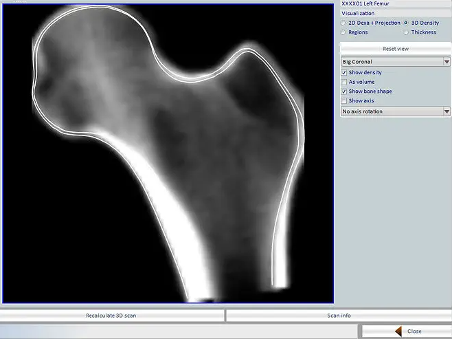
With our Medix range you obtain information on bone density in g/cm² and g/cm³. Coupled with our 3D-DXA technology you also have access to the macro and micro structure, its cortical thickness and trabecular bone.
How does it work?
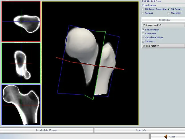
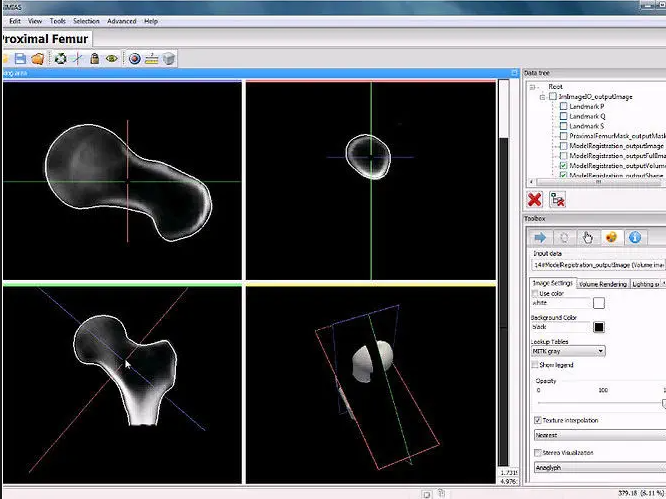
The algorithm uses a statistical model incorporating shape and density variations captured in a database of patients. This statistical model is subsequently registered onto the 2D DXA image, resulting in a 3D subject-specific reconstruction of the patient’s bone. From this 3D reconstruction can be automatically computed a set of 3D clinical parameters allowing for a better management of osteoporosis.
Integrated to the Medilink range:
- Embedded in 3D-DXA box.
- Automatic.
- Computing time 3D/2D algorithm and cortical thickness take 2 minutes
Potential
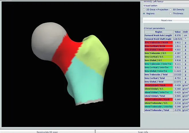
For the first time, bone health experts have access to information about bone structure which was not possible before by using DXA images, especially volumetric information:
- vBMD (volumic BMD) trabecular, cortical and global (total femur, femoral neck, intertrochanteric, greater trochanter).
- Femoral Neck Axis Length in 3D.
- Femoral Neck Shaft Angle in 3D.
- Cortical thickness 3D distribution.